-
Left ventricle (Size)
Male Normal Mildly Moderately Severely LVIDd (cm) Left ventricular internal dimension at end-diastole
4,2 - 5,8 5,9 - 6,3 6,4 - 6,8 >6,8 LVIDd (cm/m2) Left ventricular internal dimension at end-diastole
2,2 - 3,0 3,1 - 3,3 3,4 - 3,6 >3,6 LVIDs (cm) Left ventricular internal dimension at end-systole
2,5 - 4,0 4,1 - 4,3 4,4 - 4,5 >4,5 LVIDs (cm/m2) Left ventricular internal dimension at end-systole
1,3 - 2,1 2,2 - 2,3 2,4 - 2,5 >2,5 IVSd (cm) Interventricular septum thickness at end-diastole
0,6 - 1,0 1,1 - 1,3 1,4 - 1,6 >1,6 PWd (cm) Left ventricular posterior wall thickness at end-diastole
0,6 - 1,0 1,1 - 1,3 1,4 - 1,6 >1,6 RWT Relative wall thickness
0,24 - 0,42 0,43 - 0,46 0,47 - 0,51 >0,52
Recommendations for Cardiac Chamber Quantification by Echocardiography in Adults: An Update from the ASE and EACVI (2015)Female Normal Mildly Moderately Severely LVIDd (cm) Left ventricular internal dimension at end-diastole
3,8 - 5,2 5,3 - 5,6 5,7 - 6,1 >6,1 LVIDd (cm/m2) Left ventricular internal dimension at end-diastole
2,3 - 3,1 3,2 - 3,4 3,5 - 3,7 >3,7 LVIDs (cm) Left ventricular internal dimension at end-systole
2,2 - 3,5 3,6 - 3,8 3,9 - 4,1 >4,1 LVIDs (cm/m2) Left ventricular internal dimension at end-systole
1,3 - 2,1 2,2 - 2,3 2,4 - 2,6 >2,6 IVSd (cm) Interventricular septum thickness at end-diastole
0,6 - 0,9 1,0 - 1,2 1,3 - 1,5 >1,5 PWd (cm) Left ventricular posterior wall thickness at end-diastole
0,6 - 0,9 1,0 - 1,2 1,3 - 1,5 >1,5 RWT Relative wall thickness
0,22 - 0,42 0,43 - 0,47 0,48 - 0,52 >0,53 -
Left ventricle (Mass)
Male Normal Mildly Moderately Severely LV mass (g) Left ventricular mass
88 - 224 225 - 258 259 - 292 >292 LV mass (g/m2) Left ventricular mass
49 - 115 116 - 131 132 - 148 >148
Recommendations for Cardiac Chamber Quantification by Echocardiography in Adults: An Update from the ASE and EACVI (2015)Female Normal Mildly Moderately Severely LV mass (g) Left ventricular mass
67 - 162 163 - 186 187 - 210 >210 LV mass (g/m2) Left ventricular mass
43 - 95 96 - 108 109 - 121 >121 -
Left ventricle (Volume)
Male Normal Mildly Moderately Severely LVEDV (ml) Left ventricular end-diastole volume (Biplane)
62 - 150 151 - 174 175 - 200 >200 LVEDV (ml/m2) Left ventricular end-diastole volume (Biplane)
34 - 74 75 - 89 90 - 100 >100 LVESV (ml) Left ventricular end-systole volume (Biplane)
21 - 61 62 - 73 74 - 85 >85 LVESV (ml/m2) Left ventricular end-systole volume (Biplane)
11 - 31 32 - 38 39 - 45 >45
Recommendations for Cardiac Chamber Quantification by Echocardiography in Adults: An Update from the ASE and EACVI (2015)Female Normal Mildly Moderately Severely LVEDV (ml) Left ventricular end-diastole volume (Biplane)
46 - 106 107 - 120 121 - 130 >130 LVEDV (ml/m2) Left ventricular end-diastole volume (Biplane)
29 - 61 62 - 70 71 - 80 >80 LVESV (ml) Left ventricular end-systole volume (Biplane)
14 - 42 43 - 55 56 - 67 >67 LVESV (ml/m2) Left ventricular end-systole volume (Biplane)
8 - 24 25 - 32 33 - 40 >40 -
Left ventricle (Ejection fraction)
Male Normal Mildly Moderately Severely LV EF (%) Left ventricular ejection fraction (Biplane)
52 - 72 41 - 51 30 - 40 <30
Recommendations for Cardiac Chamber Quantification by Echocardiography in Adults: An Update from the ASE and EACVI (2015)Female Normal Mildly Moderately Severely LV EF (%) Left ventricular ejection fraction (Biplane)
54 - 74 41 - 53 30 - 40 <30 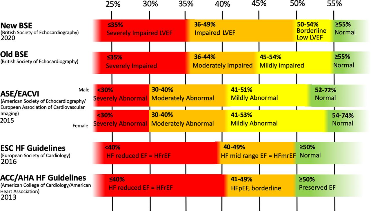 HUDSON, Sarah; PETTIT, Stephen. What is ‘normal’ left ventricular ejection fraction?. Heart, 2020.
 -
Left ventricle (Geometry)
Description of LV geometry, using at the minimum the four categories of normal geometry, concentric remodelling, and concentric and eccentric hypertrophy, should be a standard component of the echocardiography report.LV mass(g/m2) Left ventricular mass
RWT Relative wall thickness
Normal left ventricle <115 (Male) <95 (Female) <0,42 Concentric hypertrophy >115 (Male) >95 (Female) >0,42 Eccentric hypertrophy >115 (Male) >95 (Female) <0,42 Concentric remodeling <115 (Male) <95 (Female) >0,42
Recommendations for Cardiac Chamber Quantification by Echocardiography in Adults: An Update from the ASE and EACVI (2015)
Recommendations for Cardiac Chamber Quantification by Echocardiography in Adults: An Update from the ASE and EACVI (2015)LVEDV(ml/m2) Left ventricular end-diastole volume (Biplane)
LV mass(g/m2) Left ventricular mass
RWT Relative wall thickness
Normal left ventricle <75 <115 (Male) <95 (Female) 0,32-0,42 Physiological hypertrophy >75 >115 (Male) >95 (Female) 0,32-0,42 Concentric remodeling <75 <115 (Male) <95 (Female) >0,42 Eccentric remodelling >75 <115 (Male) <95 (Female) <0,32 Concentric hypertrophy <75 >115 (Male) >95 (Female) >0,42 Mixed hypertrophy >75 >115 (Male) >95 (Female) >0,42 Dilated hypertrophy >75 >115 (Male) >95 (Female) 0,32-0,42 Eccentric hypertrophy >75 >115 (Male) >95 (Female) <0,32  - The red horizontal line separates LV hypertrophy from normal LV mass.
- The black vertical line separates dilated from nondilated ventricles.
- The two oblique blue lines delimit the upper (0.42) and lower (0.32) limit of normal RWT.
- This leads to eight categories of ventricles.
- The green ellipse indicates the area of normal ventricles including physiological LV enlargement.